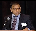
Radiofrequency Catheter Ablation for Atrial Fibrillation — Video of Kamran Rizvi, MD, FHRS

December 19, 2013
In this video from the Get in Rhythm. Stay in Rhythm.™ Atrial Fibrillation Patient Conference, Dr. Kamran Rizvi talked about radiofrequency (RF) catheter ablation, including the “stages” of afib, pre-ablation testing, and what to expect before, during, and after an RF catheter ablation.
Video watching time is approximately 20 minutes.
- Low resolution
- High resolution — YouTube
About Kamran A. Rizvi, MD, FHRS
Dr. Rizvi is a staff electrophysiologist at the Heart Hospital Baylor Plano and Co-director of the Electrophysiology Lab at the Heart Hospital Baylor Denton.
Dr. Rizvi’s undergraduate studies took place at Tulane University in New Orleans. His medical training took place at the University of Chicago Pritzker School Of Medicine. His initial training was in Internal Medicine with a highly competitive Fellowship in Cardiology at UT-Southwestern Medical School in Dallas. He completed two years of Cardiac Electrophysiology training at the University of Utah focusing on atrial fibrillation ablation. He is board certified in Internal Medicine, Cardiology and Cardiac Electrophysiology.
Dr. Rizvi has performed research on topics such as atrial fibrillation and exercise capacity.
Knowing how important this information would be to those living with atrial fibrillation, we committed to do a two-camera video shoot of the entire conference—a very expensive undertaking—in hopes that you, the afib community, will be willing to help us defray those costs through a donation (instead of us charging you for these videos, which many of you said you were willing to pay for). You can make a secure tax-deductible donation here, or click on the red Donate Now button.
Video Transcript:
Mellanie: Welcome back from break, and I hope you had a great break, and you got an opportunity to visit some of the sponsors and get some afib-healthy food. Before the break, we focused on understanding afib, and the ways to treat it with medications. For the next hour, we’ll focus on how to treat it with procedures. That’s pretty much what you do when medications don’t work. Although for some, we’re starting to look at procedures as a first line treatment rather than going through the various medications.
The first three presenters will talk about catheter ablation and managing the left atrial appendage of the heart, and then the fourth presenter will talk about surgical procedures. We will have 15 minutes each, and then we’ll ask you to hold your questions until all four have presented, and then we’ll have a panel Q&A for 35 minutes to talk about the different procedures, how to know what’s right for you, the pros and cons, and then we’ll have time for other questions and answers at the conclusion of the procedure part.
Our first procedure presenter is Dr. Kamran Rizvi. Dr. Rizvi is a staff electrophysiologist at the Heart Hospital Baylor Plano and Co-Director of the Electrophysiology Lab at the Heart Hospital Baylor Denton. He did two years of specialized atrial fibrillation ablation training at the University of Utah with someone that many in this community know quite well, and that’s Dr. Nassir Marrouche. Dr. Rizvi has published and presented research on topics such as atrial fibrillation and exercise capacity, and so we’ll also want to ask for his take on that area when we get to the Q&A. With that, let me turn it over to Dr. Rizvi.
Dr. Kamran Rizvi: Good morning everyone, I wanted to start by thanking Mellanie and her organization for being such a pivotal resource for patients with atrial fibrillation. It really is one of the best resources for anyone with afib, and I make sure to tell all my patients about the website. I’d also like to thank everyone for coming and taking the initiative to learn more about this disease on a Saturday morning.
I’m going to talk about radiofrequencyablation for atrial fibrillation. [3:01] I’m going to start with the very basics, and I apologize if this is too basic, but I just don’t want to assume anything. The heart has four chambers—there are two at the top, atrium, and two at the bottom, ventricles. The heart beats normally start at the very top of the heart—that’s the sinus node. Then, the beats progress lower down to the AV node, and then they cause the ventricles to contract, and that’s actually what produces a heartbeat. That’s God’s pacemaker, the sinus node. With atrial fibrillation, or afib, basically the top chambers of the heart are essentially having a seizure. The top chambers of the heart are beating uncontrollably fast in a very chaotic and disorganized way. As a result, the bottom chambers of the heart contract in a very chaotic and disorganized way. What’s important to realize is what triggers the afib. Where does the actual afib come from? To the best of our knowledge, the most common culprit behind atrial fibrillation is usually these structures called pulmonary veins.
[4:34] The pulmonary veins are the veins that are responsible for draining the blood that has been oxygenated in the lungs and returning that blood to the heart. There are four of them, usually. There are two that drain the right lung and there are two that drain the left lung. Some pivotal research that went on about 15 years ago showed that the triggers behind the afib, the vast majority of the time, come from the pulmonary veins, and that is very important to understand. Once you understand that, we can then begin talking about atrial fibrillation ablation. It doesn’t matter which energy modality you use, this concept is pivotal to understanding what it is we do.
[5:25] I describe afib ablation treatments—and this is very simplified, and there are a lot of caveats—I generally describe it as a Plan A, Plan B, Plan C kind of way of looking at it. The first plan, or Plan A, is medical therapy or lifestyle modifications. For example, if you have a night of heavy drinking, and you go into afib, then the therapy for that afib is going to be to avoid excessive alcohol use. If you have sleep apnea, and you’re going into afib, then I would suggest you get a sleep study, and maybe be on CPAP. Lifestyle modification is going to be our first-line option.
[6:13] Just next to that is going to be medical therapy, and all of you in this room are very familiar with beta blockers, calcium channel blockers, and antiarrhythmic drugs — those are our mainstays. But what do you do if that doesn’t work? What do you do if that fails? That’s when I talk about plan B. So, if you’ve been tried on medications, and those have not worked, and you’ve tried to adjust your lifestyle, and do all the right things, and you’re still suffering from afib, then it is reasonable to pursue an invasive strategy. And like Mellanie said, sometimes we decide to do Plan B before Plan A in certain patients. Plan B involves ablation, and that can either be with radio-frequency or it can be with cryoablation where you freeze the veins.
Plan C also can sometimes be Plan A depending on the patient, but Plan C, in my mind, is generally a pacemaker together with this thing called an AV node ablation, and essentially what we do is take over the rhythm of the heart with the pacemaker. We say, “You know what? You might have afib, it’s difficult to control, and we’re just going to take over the afib. We’re going to take over the rhythm of the heart with the pacemaker.” And that has pluses and minuses.
[7:38] Regardless of the plan we chose—Plan A, Plan B, or Plan C—regardless of the plan, it is important, as Dr. Prystowsky said, to preserve the brain and protect the brain. Regardless of the plan, we talk about blood thinners—aspirin or stronger blood thinners—regardless of Plan A, Plan B, or Plan C.
[8:05] To add to this kind of paradigm, I had the privilege of being at the University of Utah for my atrial fibrillation training where some seminal work is being done where you’re actually looking at the tissue of the heart. In Utah, they have developed these protocols where patients can get MRIs of their heart with really high resolution, and they can show us scar tissue within the heart. We know that the more scar tissue you have, the more difficult it’s going to be for that afib to be cured. I want to introduce this concept of stages of afib. I’ll use a cancer analogy, and we all know afib is not life-threatening in the way cancer is, but just for analogy purposes, people stage cancer—stage one cancer, stage two cancer—you can stage afib, as well.
[9:03] Each one of these pictures shows a left atrium, and these structures in grey are the four pulmonary veins. You can count them: one, two, three, four. These white areas and these green areas represent areas of scar. So, as you can see, some patients with a Stage 1 disease have minimal scar. Their heart is essentially normal; they just have an electrical problem. These patients do very well with ablation, and in these patients, it is reasonable to pursue ablation first-line before anything else, just right away. Stage 2 patients have more scars. Stage 3 has even more. Look at this Stage 4 patient—this patient has so much scar in the top chamber in his heart, we could ablate him, and we could do the best job in the world, and Dr. Prystowsky could do the ablation, but guess what? It’s not going to work; this patient is still going to have afib. This is a patient in whom we would want to consider just leaving that patient in afib, or even putting in a pacemaker, and saying, “Let’s just be done with it.” Does that make sense to everyone?
[10:16] What do we do with radiofrequency ablation? Radiofrequency ablation is the most common way we ablate afib, and it gets back to that pulmonary vein concept. So, essentially what we try to do is take this catheter—this catheter in this picture has an irritated tip, so there’s some saline shooting out of the tip of the catheter—this catheter is capable of delivering radiofrequency energy and essentially cauterizing the heart. Our goal is to actually build a fence, build a wall around these pulmonary veins. And we say, “Hey, this pulmonary vein may be triggering the afib with abnormal firing, and what we’re going to do is we are going to block those signals.” So, those signals are going to be trapped, and they cannot allow the heart go into afib. We’re going to try to fix the problem at the source.
[11:17] This is an example of a typical ablation lesion set, and I know it looks like a cartoon that a six-year-old might draw, but there’s actually a lot of technology that goes into this. What this picture represents is the left atrium, and there are four pulmonary veins pictured here: one is pink, one is yellow, one is green, and one is light blue. Those are the four pulmonary veins. These red circles represent where we ablated. So, that is where we delivered energy. If you can picture with me, we basically went around each one of these pulmonary veins in a circular fashion in an effort to isolate those veins, and again build a fence around those veins. This is an ablation I did about three months ago; this patient has not had any more afib since the ablation. This is a typical lesion set that we do when we talk about afib ablation, so each one of these red dots is where I delivered energy in an effort to isolate the veins. The other name for afib ablation is pulmonary vein isolation. So, those are synonymous.
[12:42] If we decide to pursue an afib ablation with radiofrequency, there are certain steps and procedures involved, and I do want to take a moment to go through those. The first is, in most cases, we would ask you to have an echo, and this is not an echo on the surface of the heart. This is an echo that actually goes into the food tube, and that’s important because the food tube sits right next to the left atrium. So, we want to make sure there are no clots sitting in your heart before we go in. Every patient that gets an afib ablation typically gets a transesophageal echo to rule out a preexisting clot in the heart because the last thing we want to do is dislodge that clot and cause a potential stroke. That’s one test to get prior to an ablation.
The other one is a CT scan or a cardiac MRI. That test is done to really define the anatomy of the left atrium. We want to make sure we have a good understanding of what the anatomy looks like before we go in. Most people have four veins, but some people have three veins, some people have five veins. There’s, you know, an infinite number of permutations of possible pulmonary vein anatomy, and the CT scan helps to really define that for us. The MRI additionally can offer additional information regarding the scar in the atrium, like I showed you before, but not every center can do that quite yet. Most places are still doing CT scans.
[14:28] In general with the ablation, we ask you not to eat anything the night prior, and that’s important because the anesthesiologist wants you to have an empty stomach because the medicines that they give may well cause you to have some nausea. They want to make sure there’s no risk of vomiting. So, an empty stomach; nothing to eat the night prior. We also usually recommend you stop a lot of your heart-related medicines prior, and what I mean by that are two things in particular. Number one is antiarrhythmic medications. If you’re on an antiarrhythmic medication, such as Flecainide, Rythmol, Betapace, or Amiodarone, we recommend you typically stop those because we want those to wash out of your system; we want to see your heart as it is without the interference of any medication. That’s important because if we’re going to be able to tell if the vein is isolated or not, we don’t want any meds that would potentially obfuscate that determination—we want to see the heart without the interference of antiarrhythmic medication.
[15:34] Number two, blood thinners. Usually, prior to an ablation, we would ask you to stop your blood thinners, and there are some doctors that will ask you to continue your blood thinner; that is something that is really dependent on the doctor.
[15:48] The ablation typically takes about three hours with the range of two to four hours. We typically use general anesthesia, although in Utah where I trained, we did not use general anesthesia; most places will use general anesthesia. We put these catheters into the heart, which are these thin, floppy tubes—we put them into the heart and that can help us do the actual ablation. Post-procedure, we remove all the catheters from the heart so there’s no hardware left; everything is out. We hold pressure over the veins, we ask you to have a period of bed rest. If all goes well, you go home the next day.
[16:30] What is the success rate for ablation? Most folks nowadays are quoting success rate for a single procedure of about 70 to 80 percent. Now, there are a lot of caveats with that number. That is a success rate with a single procedure with afib that is paroxysmal or intermittent. So, if you have more advanced afib, that number is not going to be as high. If you have intermittent afib, 70% to 80% is a reasonable number. Bear in mind that that is a very strict definition of success. That is defined as less than 30 seconds of afib over the next year. So, if you have 31 seconds that is considered a failure. I think we would all agree that’s still a success, but this is the strict definition of success. I would argue partial success is a lot higher.
[17:28] Major complication rate is approximately 1%–2%, with a total rate of about 3%–5%. And most of that 3%–5% is what I call groin-related, access site-related. You can have some bleeding down in your groins, it could turn all the colors of the rainbow, but it will get better. You’re no worse for the wear, it’ll get better, you’ll be fine. Dangerous risk is around 1%–2%, and those include fluid in the sac around the heart, which happens about one percent of the time, and we can always manage that conservatively, as well as the potential of a stroke, which is actually very, very low.
[18:12] I’m going to move a little faster here through the slides since I am running out of time. I do want to mention for the folks that live in the Dallas/Fort Worth area, we are doing some very exciting things at the Heart Hospital. We are involved in many research projects with regards to atrial fibrillation. My partner, Dr. Deville, is also involved in a lot of these projects, and we think we’ve got some neat things that could potentially change the face of atrial fibrillation ablation.
[18:43] We also use a remote navigation system. [18:49] These are some of the trials that we take part in—the CABANA Trial, and some other trials that are potentially going to change the game with respect to atrial fibrillation ablation.
[18:59] Thank you for your attention. I’m so excited to see this crowd. If you have any questions, feel free to come up to me afterwards.
Mellanie: Thanks, Dr. Rizvi.
